Joint Preservation Procedures

Joint Preservation Procedures

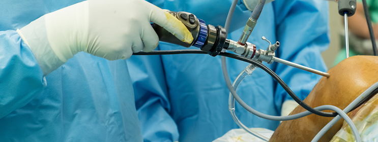
Joint preservation techniques are used in patients with cartilage defects to preserve the joints and to restore the function.
Articular cartilage is the cartilage that covers the bony surface of joints. It has a smooth surface which allows the bones of the knee joint to slide over each other with very little friction. Articular cartilage is often damaged by injury or normal wear and tear. Articular cartilage, when damaged or worn away, the affected joint becomes painful, stiff, and has limited range of motion. As the articular cartilage has limited ability to heal by itself, surgical repair may be required to stimulate the growth of new cartilage. Articular cartilage restoration relieves pain, improve function and can delay or prevent the onset of arthritis in the joint.

  • Microfracture surgery: Microfracture surgery is appropriate for patients having single lesion and healthy subchondral bone. Microfracture can be done using an arthroscope a long, thin device with a tiny camera attached at the end to see inside your knee. Your surgeon uses a small pointed tool called an awl to make very small holes, microfractures, in the bone underlying the cartilage called subchondral bone. This stimulates the healing process by increasing the blood flow to the surface which brings in new cells that build new cartilage.
  • Drilling: This technique involves use of a surgical drill or a fine wire to make multiple small holes through the damaged area to penetrate the subchondral bone. This generates a healing response within the defect. As the heat produced during drilling can cause injury to some of the tissues, it is considered less accurate than microfracture.
  • Abrasion arthroplasty: Abrasion arthroplasty can be done using an arthroscope. This technique is similar to drilling, rather than drills or wires, high speed burrs are used to remove the damaged cartilage and to penetrate the subchondral bone.
  • Autologous chondrocyte implantation: Autologous chondrocyte implantation (ACI) is a two-step procedure; first step is an arthroscopic surgery to remove the tissue containing healthy cartilage cells from an area of the bone that does not carry weight. These cells are cultured and multiplied in laboratory over a 3- to 5- week period. In second step, the newly grown cells are implanted through an open surgical procedure, or arthrotomy. During this surgery a periosteum, layer of bone-lining tissue is sewn over the area of damaged cartilage. Once the area is sealed with fibrin glue the cultured cartilage cells are injected underneath the periosteal cover.

Treatment's

Contact Us


Top